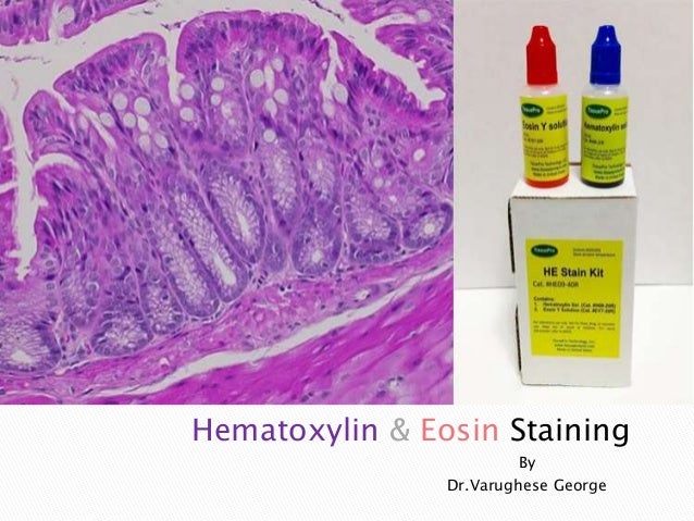
Types Of Hematoxylin Dyes
Essentially the hematoxylin component stains the cell nuclei blue-black showing good intranuclear detail while the eosin stains cell cytoplasm and most connective tissue fibers in varying shades and intensities of pink orange and red. The samples are formalin-fixed paraffin-embedded sections or frozen sections.
 An Intro To Hematoxylin Staining Protocol Hematein Formation Leica Biosystems
This stain produces colors different tissue structures which would otherwise be transparent so that you can get a detailed view of the tissue.
Types of hematoxylin dyes. Harris hematoxylin is routinely used as the preparation is easy and has less ripening time. An acidic eosin counterstains the basic elements such as RBCs cytoplasm muscle and collagen in varying intensities of pink orange and red. Phosphotungstic acid is used as mordant.
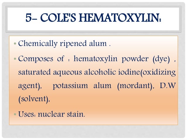
Considering all of its versatility in routine and rare variations of Hematoxylin staining it can stain the following. Harriss Mayers Carazzis and Gills. What are the types of hematoxylins.
Weigerts Hematoxylin an iron hematoxylin dye is used to stain the nuclei. Eosin Yellow Y is the most commonly used stain in histopathology laboratory. Can be prepared using hematein no oxidation required.
Stains in shades of blue and red. There are typically three types of HE stains. Hematoxylin not a dye itself produces the blue Hematin via an oxidation reaction with nuclear histones causing nuclei to show blue.
The hematoxylin stains cell nuclei blue and eosin stains the extracellular matrix and cytoplasm pink with other structures taking on different shades hues and combinations of these colors. The rest of the cell. Use as a textile dye.
Routine stain for nervous tissue also used to stain muscle striations and fibrin. This stain binds preferably to the nucleus because it is more basophilic. These differ in part according to the oxidizing agent added eg.
Nuclei mitotic structure mitochondria mucin hemoglobin elastic fibers muscle. Probably the Hematoxylin-Eosin staining HE. Regressive stain with staining time of 1-16 hours at room temperature and 1-2 hours at 60ºC.
There are a few different types of haematoxylin. Which type of hematoxylin is routinely used and what is staining method. 1 Extraction and purification.
Alum hematoxylin Purpleblue coloration Alum-hematoxylin stains nuclei various shades of purpleblue Specific staining Non-specific staining may also occur o Cytoplasm o Mucin. Hematoxylin is oxidized naturally can also be oxidized using potassium permanganate. Its actually the oxidation product of hematoxylin hematin which constitutes the dye and not actually haematoxylin itself.
To produce a functional dye hematoxylin is oxidized to hematein and subsequently is bound to one of several metal ions including aluminum Al 3 iron Fe 3 and chromium Cr 3. This dye is resistant to decolorization by acidic staining solutions. The stain shows the general layout and distribution of cells and provides a general overview of a tissue samples structure.
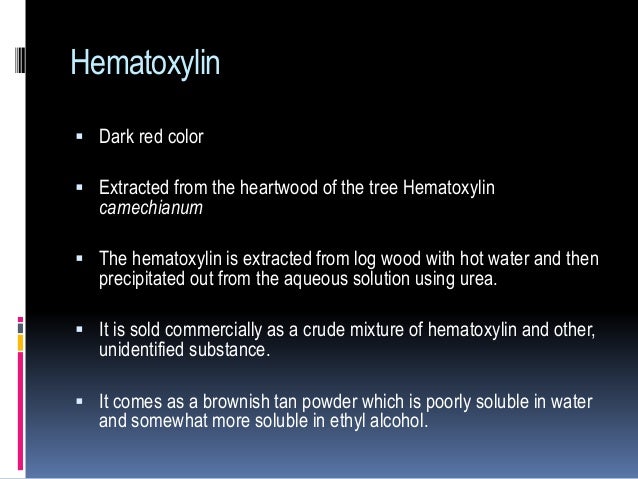
Hematoxylin the most commonly used nuclear dye most commonly used natural dye extracted from heartwood of the logwood tree which is native to Central America made in USA. Mallorys phosphotungstic acid hematoxylin. Reagents Distilled water Alum hematoxylin Acid alcohol Scotts tap water Eosin dye.
Use as a histologic stain. Hematoxylin a basic dye imparts blue-purple contrast on basophilic structures primarily those containing nucleic acid moeties such as chromtatin ribosomes and cytoplasmic regions rich in RNA. Oxidized hematoxylin is combined with aluminum ions to form an active metaldye complex that stains the nuclei of mammalian cells blue by binding to lysine residues on nuclear histones.
This allows the nuclei to be stained with a dark bluish or purple color due to its interaction with the dye Hematoxylin 091922 while the cytoplasmic components to stain pink due to Eosin interaction. Mercuric oxide Harriss or sodium iodate Gills. A metallic ion bound to a dye that is involved in the binding of the dye to tissue is referred to as a mordant.
The main difference between this dye and hematoxylin is that Nuclear fast red stains nucleic acids red in only about 5 minutes. Anionic dyes are important for staining cytoplasm extracellular structures. Biebrich scarlet-acid fuschin solution stains all the acidic tissues such as the cytoplasm muscle and collagen.
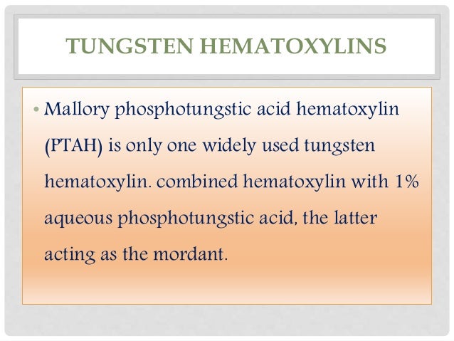
This is in contrast to other nuclear stains that label the nucleic acids. TUNGSTUN HEMATOXYLIN Mallory phosphotungstic acid hematoxylin PTAH is an example. Progressive staining occurs when the hematoxylin is added to the tissue without being followed by a differentiator to remove excess dye.
Progressive modified progressive and regressive. Use as a writing and drawing ink. As its name suggests HE stain makes use of a combination of two dyes haematoxylin and eosin.
Nuclear fast red also called Kernechtrot dye is another nuclear stain. Hematoxylin Hematein complexed with Al3 is the most common form of hematoxylin used for nuclear staining. PREPARATION OF HEMATOXYLIN STAIN.
2 Use as a histologic stain. Hematoxylin and eosin Hematoxylin and eosin HE is the most widely used stain in histology and allows localization of nuclei and extracellular proteins. Various types of Eosin stains are available like Pink Red Orange and Yellow.
Haematoxylin and Eosin For routine examination haematoxylin and eosin HE is the stain of choice.
 H E And Special Staining Histology Services Research Cro Custom Services
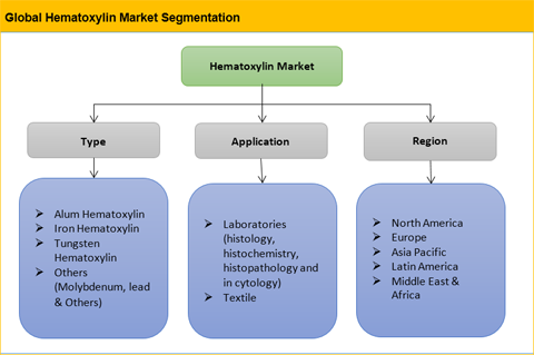 Hematoxylin Market Size Share Trend And Forecast To 2025
 Hematoxylin Eosin Staining On Mouse Sternum Stain Mouse Presents
 Histology Hematoxylin Eosin Staining Of A Native Skin Biopsy B Download Scientific Diagram
 Hematoxylin An Overview Sciencedirect Topics
H E Staining Part 2
 Hematoxylin Amp Eosinstaining
 Hematoxylin And Eosin Staining
 Application Of Hematoxylin Reagent For Sperm Cell Separation In Sexual Crime Evidence Sciencedirect
 Histology Hematoxylin Eosin Staining Showing Heterotopic Gastric Download Scientific Diagram
 Hematoxylin And Eosin Stain Medical Videos Stain H E Stain
 An Intro To Hematoxylin Staining Protocol Hematein Formation Leica Biosystems
 Image Result For Pseudo Stratified Columnar Epithelial Cells Cell Tie Dye Skirt Tie Dye
 Hematoxylin And Eosin
 Iron Hematoxylin Staining Staining Microbe Notes
 Hematoxylin And Eosin
 Spectral Characterization Of Hematoxylin Eosin H E And Nearinfrared Download Scientific Diagram