What Causes A Hemangioma On The Spleen
Rarely growing hemangioma can cause signs and symptoms that may require treatment including pain in the upper right quadrant of the abdomen abdominal bloating or nausea. A hemangioma is made up of extra blood vessels that group together into a dense clump.

Occasionally a hemangioma can break down and develop a sore.

What causes a hemangioma on the spleen. Spleen trauma can be very serious because it can cause a potential spleen rupture. Traumatic physical injuries are another cause of lesions on the spleen. The female hormone estrogen which increases during pregnancy is believed to cause some liver hemangiomas to grow larger.
The most common benign primary neoplasm of the spleen is a hemangioma. Injuries to the abdominal area caused by fighting or an accident can injure the spleen. Dear JTS hemangiomas of the spleen are beyond the area of expertise of thiss Forum.
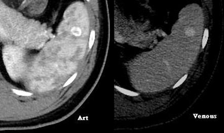
Which cancers present with a mass on the spleen. These tumors form by proliferation of vascular channels ranging from small capillaries to large cavernous types. After a spleen ruptures a person can bleed internally which can be life-threatening.
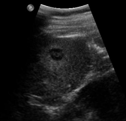
1 Focal echogenic lesions in the spleen in sickle cell disease. However spontaneous rupture has been reported to occur in as many as 25 of this patient population1 Treatment most often consists of splenectomy. Hemangiosarcoma is cancer of the vascular endothelium or the blood vessel walls.
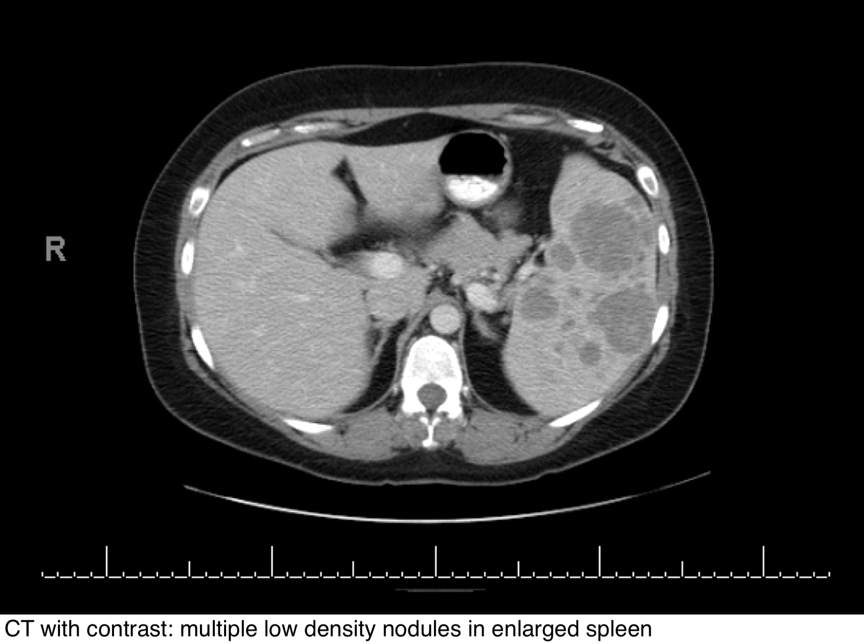
Diffuse hemangioma of the spleen occurred in a 59-year-old man. What causes the vessels to clump isnt known. It is aggressive and spreads fast to other parts of the body especially the lungs.
Benign growths usually imply that the organ affected will not require removal but because of the concentration of blood vessels located within the spleen a splenectomy likely be recommended by the physician. Other cancers such as breast cancer melanoma and lung cancer can spread to the spleen. Cancer in the spleen is usually caused by lymphomas and leukemias.
The most likely cause of canine hemangiosarcoma is genetic predisposition which was observed in studies to be likely responsible for the occurrence of this disease and it almost always occurs in. Case reports have associated LCA with various other conditions including portal hypertension Crohns disease Gaucher disease lymphoma aplastic anemia colon cancer pancreatic cancer lung cancer and myelodysplastic syndrome. It often has a latent clinical picture.
Besides abdominal pain LCA may cause an enlarged spleen splenomegaly anemia or thrombocytopenia. It commonly arises from the spleen the liver and the skin. It often has a latent clinical picture.
A spleen hemangioma is the most common type of benign mass that might develop on the spleen. Although most angiomas do not cause problems some can grow rapidly and cause pain andor bleeding. A hemangioma is a slow-growing neoplasm consisting of an overgrowth of new blood vessels and it is found most often when a patient is being screened for another illness.
They are usually incidental findings during the investigation of other problems. It accounts for 02 to 3 percent of all canine tumors with a mean age at diagnosis of 9 to12 years1 Hemangiosarcoma most commonly affects the spleen and heart of golden retrievers Labrador retrievers and German shepherds. Was diagnosed with a mass on spleen possibly a hemangioma but indeterminate.
This report reviews an 8-year experience with splenic hemangioma at Mayo Clinic. The cancer cells invade the endothelial cells lining the walls of the blood vessels in the affected organ. Splenic hemangioma is a rare disorder but remains the most common benign neoplasm of the spleen.
Most hemangiomas are asymptomatic and incidentally discovered. Splenic hemangioma is a rare disorder but remains the most common benign neoplasm of the spleen. Kasabach-Merritt syndrome characterized by hemangiomatosis thrombocytopenia and intravascular coagulation is a rare syndrome resulting from sequestration of red blood cells and platelets and consumption of clotting factors in the hemangiomas typically seen in early infancy.
However spontaneous rupture has been reported to occur in as many as 25 of this patient population. Splenic hemangiomas also known as splenic venous malformations splenic cavernous malformations or splenic slow flow venous malformations while being rare lesions are considered the second commonest focal lesion involving the spleen after simple splenic cysts 512 and the most common primary benign neoplasm of the spleen 6. Hemangiomas occur more often in babies who are female white and born prematurely.
Hemangiosarcoma HSA arises from blood vessels. Hemangiomas are common abnormalities of the abdominal organs. This nasty cancer affects the skin heart liver or spleen.
Big clues that your pet may have this cancer include signs of vague lethargy or waxing and waning weakness. Occasionally it occurs in other parts including the heart. 1 Treatment most often consists of splenectomy.
This report reviews an 8-year experience with splenic hemangioma at Mayo Clinic. Ed Friedlander answered 43 years experience Pathology. Hemangiosarcoma is a soft tissue tumor sarcoma that arises out of blood vessels the arteries or veins.
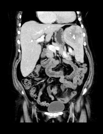
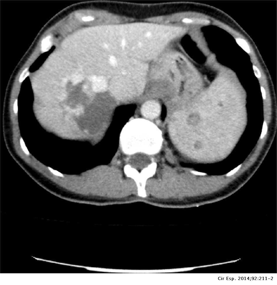
5 Isolated hemangiomas are seen most commonly although occasionally multiple may be present. The presenting features were a dull ache and heaviness in the left upper quadrant for 3 weeks severe left-sided pain and fever. They are usually found incidentally and have imaging appearances similar to hepatic hemangiomas.
Hemangioma of spleen with spontaneous extra-peritoneal rupture with associated splenic tuberculosis an unusual presentation Australasian Radiology Vol.


Http Pdf Posterng Netkey At Download Index Php Module Get Pdf By Id Poster Id 109214